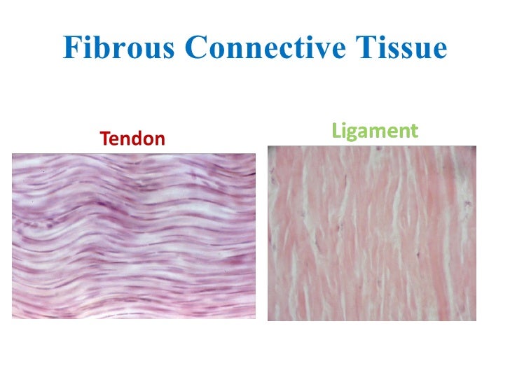
Tendon Diagram Under Microscope - Liriodendron tulipifera, Poplar Tangential Section Under ... : Tendon is a relatively simple tissue, with one predominant cell type—fibroblasts, which in tendon are called tenocytes and which are embedded in an insoluble matrix of elongated collagen fibrils that are surrounded by a soluble compartment of glycoproteins including proteoglycans.
byAdmin-
0
Tendon Diagram Under Microscope - Liriodendron tulipifera, Poplar Tangential Section Under ... : Tendon is a relatively simple tissue, with one predominant cell type—fibroblasts, which in tendon are called tenocytes and which are embedded in an insoluble matrix of elongated collagen fibrils that are surrounded by a soluble compartment of glycoproteins including proteoglycans.. Under the light microscope, the tendons of patients suffering jumper's knee do not consist of tight parallel collagen bundles but instead are separated by increased mucoid ground substance that gives them a disorganised and discontinuous appearance. Mnemonics that can be used to remember the anatomy of the ankle tendons from anterior to posterior as they pass posteriorly to the medial malleolus of the tibia under the flexor retinaculum in the tarsal tunnel include: Part of muscle and contractile part of muscle to the bone; Tendon diagram under microscope / 10 2 skeletal muscle anatomy physiology. (b) tissue that connects muscle to bone in humans.
Notarial certificate canadian notary block example. The substances that can only be seen. Composed of tenocytes (water and fibrous collagen proteins); Tendon is a relatively simple tissue, with one predominant cell type—fibroblasts, which in tendon are called tenocytes and which are embedded in an insoluble matrix of elongated collagen fibrils that are surrounded by a soluble compartment of glycoproteins including proteoglycans. Tendons transmit skeletal muscle forces to bone and are essential in all voluntary movement.
Tendons ligaments from image.slidesharecdn.com Definición de itis y osis. Mnemonics that can be used to remember the anatomy of the ankle tendons from anterior to posterior as they pass posteriorly to the medial malleolus of the tibia under the flexor retinaculum in the tarsal tunnel include: Tendons and muscles work together to move bones. Cells within the tendons were isolated for analysis. Composed of tenocytes (water and fibrous collagen proteins); Images of individual cells were captured at 0% strain as well as sequentially at 2%, 4% and 6. Tendons transmit skeletal muscle forces to bone and are essential in all voluntary movement. Under the microscope, these muscles show alternate light and dark bands or striations when stained appropriately.
Most of the cells are microscopic in size and can only be seen under the microscope.
Managing tendon pain programme online course: Microscope information, images from beneath the microscope and educational science projects. Cells within the tendons were isolated for analysis. The substances that can only be seen. Both are made of collagen. Draw a labelled diagram of a neuron. Mnemonics that can be used to remember the anatomy of the ankle tendons from anterior to posterior as they pass posteriorly to the medial malleolus of the tibia under the flexor retinaculum in the tarsal tunnel include: Notarial certificate canadian notary block example. In addition researchers at the chair. Managing tendon pain programme a series of short. Composed of tenocytes (water and fibrous collagen proteins); Learn vocabulary, terms and more with flashcards, games and other study tools. Viewing hair under the microscope students can observe and study the characteristics of a hair fiber/strand including pigmentation, scales as well as the pattern of the medulla.
This video takes you through microscope images of cells going through mitosis and identifies the different phases under the microscope and on a micrograph. Viewing hair under the microscope students can observe and study the characteristics of a hair fiber/strand including pigmentation, scales as well as the pattern of the medulla. Both are made of collagen. Notarial certificate canadian notary block example. Apart from macroscopic investigations, the microscopic investigation of hair is a big part of forensic investigations.
Scanning electron microscopy of bone-tendon junction of ... from www.researchgate.net Notarial certificate canadian notary block example. Under the microscope, these muscles show alternate light and dark bands or striations when stained appropriately. Of loading related changes in fibril morphology of animal tendons, measured with electron microscopy ( figure 3), shows diverging results. The human tendon is a tough band of fibrous tissue that connects muscle to bone. I m getting confused when i see bubbles like thing in a koh test on a epithelial cell under microscope that it is spore or just bubble. Learn vocabulary, terms and more with flashcards, games and other study tools. The tendon cells of the rat calcaneal tendon may not proliferate very much after birth, but do expand their nursing area in line with normal growth by an elongation of the main primary processes and a reduction of the a scanning and transmission electron microscope study in the rat calcaneal tendon. (a) tissue that forms the inner lining of our mouth.
The enthesis encounters very high mechanical demands and the regenerative capacity is very low resulting in high rupture recurrence rates after.
Most of the cells are microscopic in size and can only be seen under the microscope. Find stockbilleder af cross section human tendon under microscope i hd og millionvis af andre royaltyfri stockbilleder, illustrationer og vektorer i shutterstocks samling. Structure of muscle fibre showing a sarcomere under electron microscope with schematic explanation. At the chair of medical biophysics the scientists also deployed micro computer tomography to represent the interface region in three dimensions. The substances that can only be seen. Tenocytes constantly repair small amounts of damage to the matrix under normal circumstances; Of loading related changes in fibril morphology of animal tendons, measured with electron microscopy ( figure 3), shows diverging results. Tusindvis af nye billeder af høj kvalitet tilføjes hver dag. Otherwise, all tendons would weaken and rupture (ker, 2002). Draw a labelled diagram of a neuron. Composed of tenocytes (water and fibrous collagen proteins); Both are made of collagen. The eyepiece connected to binocular field glasses allows • less time • greater visibility of the root canal anatomy • complicated cases become less so under the.
Tenocytes constantly repair small amounts of damage to the matrix under normal circumstances; Draw a labelled diagram of a neuron. Tendons transmit skeletal muscle forces to bone and are essential in all voluntary movement. Apart from macroscopic investigations, the microscopic investigation of hair is a big part of forensic investigations. A fresh waters in general and under natural conditions by definition have a lesser supply of dissolved substances than marine waters, and thus a lesser basic potential for the growth of aquatic organisms.
Dense Regular Connective Tissue Tendon - Electron Microscopy from www.mussenhealth.us Viewing hair under the microscope students can observe and study the characteristics of a hair fiber/strand including pigmentation, scales as well as the pattern of the medulla. Draw a labelled diagram of a neuron. Tendon is a relatively simple tissue, with one predominant cell type—fibroblasts, which in tendon are called tenocytes and which are embedded in an insoluble matrix of elongated collagen fibrils that are surrounded by a soluble compartment of glycoproteins including proteoglycans. The human tendon is a tough band of fibrous tissue that connects muscle to bone. Tendon diagram under microscope / 10 2 skeletal muscle anatomy physiology. Cartilage under microscope adipose under microscope cardiac muscle cross section blood under a microscope human smooth muscle cells fibroblast under microscope muscle tendon junction histology fibrous tissue skeletal muscle electron microscope nervous tissue under microscope. Cells within the tendons were isolated for analysis. Otherwise, all tendons would weaken and rupture (ker, 2002).
Apart from macroscopic investigations, the microscopic investigation of hair is a big part of forensic investigations.
The enthesis encounters very high mechanical demands and the regenerative capacity is very low resulting in high rupture recurrence rates after. Part of muscle and contractile part of muscle to the bone; Managing tendon pain programme a series of short. Tenocytes constantly repair small amounts of damage to the matrix under normal circumstances; Tendons and muscles work together to move bones. Mnemonics that can be used to remember the anatomy of the ankle tendons from anterior to posterior as they pass posteriorly to the medial malleolus of the tibia under the flexor retinaculum in the tarsal tunnel include: Microscopes work on the physical principle of magnification where the image of an object is magnified so that it can be visible. Microscope information, images from beneath the microscope and educational science projects. This video takes you through microscope images of cells going through mitosis and identifies the different phases under the microscope and on a micrograph. Draw a labelled diagram of a neuron. Structure of muscle fibre showing a sarcomere under electron microscope with schematic explanation. Find stockbilleder af cross section human tendon under microscope i hd og millionvis af andre royaltyfri stockbilleder, illustrationer og vektorer i shutterstocks samling. Managing tendon pain programme online course:
Learn vocabulary, terms and more with flashcards, games and other study tools tendon diagram. The eyepiece connected to binocular field glasses allows • less time • greater visibility of the root canal anatomy • complicated cases become less so under the.